IBS vs IBD: a one-letter difference that changes everything.
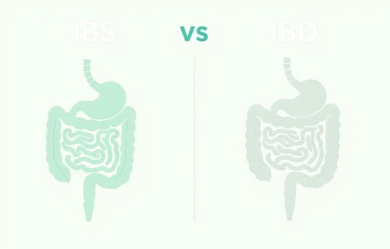
One letter. IBS and IBD look alike on the page and feel alike at the start. The plan after diagnosis could not be more different.
IBS is irritable bowel syndrome. It is one of the disorders of gut-brain interaction (DGBI). The gut looks normal on every test. The wiring between gut and brain is the issue. IBD is inflammatory bowel disease, which covers Crohn's disease and ulcerative colitis. That one is structural. Real inflammation. Real tissue damage that a camera and a biopsy can see.
If a clinician calls the wrong one, the next year of your care points in the wrong direction. So the read-out you actually need is which of these two stories your gut is telling.
What IBS is
IBS is a pattern, not a wound. The Rome V criteria (2026) define it by recurrent abdominal pain or discomfort linked to defecation, change in stool frequency, or change in stool form, present at least three days per month over the prior three months. Colonoscopy is normal. Bloodwork is normal. Imaging is normal.
What is actually happening is a miscommunication along the gut-brain axis. The intestines move, sense, and signal in ways that produce real symptoms without producing visible damage. Visceral hypersensitivity, altered motility, central sensitization, and stress-driven inputs all play a role. The gut is firing alarms when it should be quiet. That is the wiring problem.
IBS is real. The pain is real. The bloating is real. There is nothing on the scope camera to point to, and that is part of the definition, not a failure of the test.
What IBD is
IBD is damage. The lining of the bowel is inflamed, ulcerated, or eroded, and a pathologist can see it under a microscope. Crohn's disease can hit any segment of the gut from mouth to anus, often with patchy involvement and full-thickness inflammation. Ulcerative colitis is confined to the colon and rectum and tends to be continuous starting at the rectum.
The cause is immune-driven. The body's own inflammatory response attacks the gut wall, and over time that produces strictures, fistulas, abscesses, ulcers, and an elevated lifetime risk of colon cancer if poorly controlled. Treatment is also immune-driven. Mesalamine, steroids in flares, immunomodulators, and biologic drugs that quiet the inflammation are the backbone of care. The American Gastroenterological Association (Singh 2021) and the American College of Gastroenterology (Lichtenstein 2018, Rubin 2019) publish the treatment frameworks.
IBD is not something you manage with diet alone. Diet helps. Diet does not put out the fire.
Where they look identical
Early on, the overlap is the trap. Both can present with abdominal pain that comes in waves. Both can cause diarrhea, sometimes urgent. Both can flare with stress. Both can leave you bloated, tired, and afraid of meals. Cramping after eating is common to both. So is a sense that your gut runs the calendar.
If the only inputs are symptom history and a physical exam, the two can blur. That is exactly why workup matters. The history alone is not enough.
Where they part: the red flags
Certain features do not fit IBS and require a structural workup. These are the signs that the story is not just wiring.
None of these red flags prove IBD on their own. They mean the differential is wider than IBS and the right next step is testing, not reassurance.
The tests that split them
Two tests do most of the work.
Fecal calprotectin is a stool test that measures a protein released by white blood cells in the gut wall when there is inflammation. It is cheap, noninvasive, and reasonably accurate at separating IBS from IBD. The American Gastroenterological Association guideline on functional diarrhea and IBS (Smalley 2019) endorses calprotectin as a first-tier test in patients without alarm features who need IBS confirmed. Low calprotectin is reassuring. Elevated calprotectin pushes the workup toward IBD or another inflammatory cause.
Colonoscopy with biopsy is the structural test. A gastroenterologist passes a camera through the colon and the end of the small intestine, looks at the lining directly, and takes tissue samples. In IBS, the scope is normal and the biopsies are normal. In IBD, the scope shows inflammation, ulceration, or characteristic patchy or continuous disease, and the biopsies confirm it under the microscope. Mayo Clinic and ACG reviews (Lichtenstein 2018) frame colonoscopy with biopsy as the diagnostic anchor for IBD.
Other tests fill in around those two. CBC for anemia. CRP for inflammation. CT or MR enterography to map small bowel disease in suspected Crohn's. The point is that IBD has objective evidence. IBS does not need that evidence because the definition is symptom-based with a normal structural workup.
Side by side
| IBS | IBD | |
|---|---|---|
| Mechanism | Disorder of gut-brain interaction. Wiring problem. | Immune-driven inflammation. Structural damage to the gut wall. |
| Colonoscopy appearance | Normal lining. Normal biopsies. | Inflammation, ulcers, or characteristic patchy or continuous disease. Abnormal biopsies. |
| Fecal calprotectin | Typically low. | Typically elevated, often markedly so during flares. |
| Blood in stool | Not a feature. If present, requires workup. | Common, especially in ulcerative colitis. |
| Weight loss, fevers, night sweats | Not a feature. | Common in active disease. |
| Treatment direction | Diet adjustment, fiber strategy, gut-brain therapies, neuromodulators, daily symptom tracking. | Mesalamine, steroids in flares, immunomodulators, biologic drugs. Surgery in select cases. |
What to do
- Track symptoms daily for at least 30 days. Note stool form, pain timing, food, sleep, and stress. Pattern is the most useful input you can bring to a gastroenterologist.
- Identify your red flags honestly. Blood in stool, weight loss, fevers, night sweats, anemia, a family history of IBD or colon cancer, or new symptoms after age 50 all change the plan.
- Ask your clinician for a fecal calprotectin test if IBS has not been confirmed. It is a cheap stool test that helps separate wiring from inflammation.
- Ask about colonoscopy with biopsy if red flags are present or if calprotectin is elevated. This is the test that confirms or rules out IBD.
- Follow up with a gastroenterologist for diagnosis and an ongoing plan. The label drives the treatment. Get the label right.
One letter. Two completely different roads. Know which one you are on.